Search
×
Search
×
Search
×
Salud familiar e infantil
Compartir
Minutos de lectura
El ultrasonido usa ondas sonoras que producen una imagen del interior de una parte de su cuerpo. El ultrasonido se usa durante el embarazo para monitorear el crecimiento de su bebé y detectar anormalidades físicas.
Usted se reunirá con el obstetra o el auxiliar de ecografía que llevará a cabo el procedimiento para discutir su tratamiento. Puede ser distinto del procedimiento descrito aquí, ya que estará diseñado de acuerdo con sus propias necesidades. Los detalles del procedimiento también pueden variar de un país a otro.
ÍNDICE DEL ARTÍCULO
El ultrasonido utiliza ondas sonoras de alta frecuencia y sus ecos para crear imágenes en movimiento tridimensionales (3D) o en cuatro dimensiones (4D) de su bebé en crecimiento. Las imágenes (exámenes exploratorios) se ven en negro, blanco y gris y se visualizan en la pantalla de un monitor que usted podrá ver durante el procedimiento.
Los ultrasonidos en el embarazo generalmente son realizados por un obstetra o un auxiliar de ecografía. Un obstetra es un médico que se especializa en aspectos relacionados con el embarazo y el parto. Un auxiliar de ecografía es un técnico que está especialmente capacitado para tomar ecografías.
Hay diferentes motivos para realizar los ultrasonidos durante diferentes etapas del embarazo. Durante el embarazo, es muy probable que le ofrezcan al menos dos exploraciones – una ‘ecografía para determinar la edad gestacional’ destinada a comprobar la fecha de parto, y una ‘exploración de anomalía fetal’ para comprobar que su bebé se está desarrollando normalmente. Se le podrían indicar exploraciones adicionales si tiene un mayor riesgo de problemas de salud o tiene antecedentes familiares de ciertas condiciones médicas que puedan afectar su embarazo.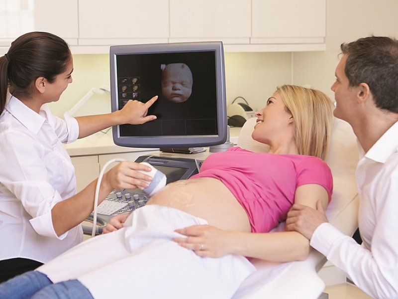
Ecografía para determinar la edad gestacional
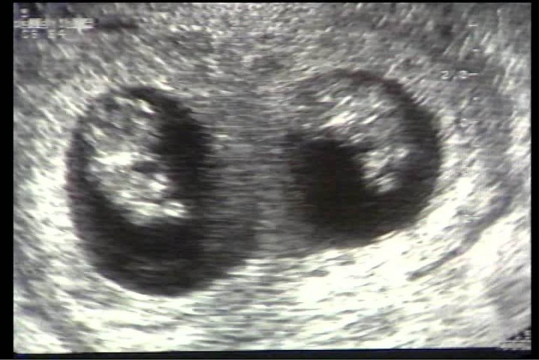
Generalmente se le ofrecerá un ultrasonido entre las semanas 10 y 14 del embarazo para confirmar cuándo nacerá su bebé. Conocer la edad gestacional de su bebé puede ayudar a monitorear los eventos importantes de su embarazo. Este examen exploratorio también le dirá si usted está esperando más de un bebé. Si le practican el examen exploratorio entre las semanas 11 y 13 del embarazo, también se puede determinar si su bebé padece de síndrome de Down.
Examen exploratorio de anomalías fetales
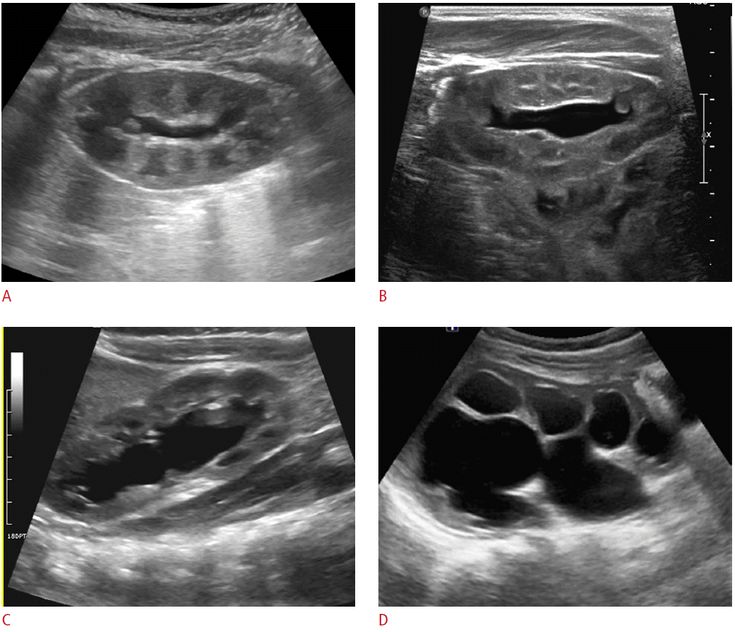
Generalmente se le ofrecerá un ultrasonido entre las semanas 18 y 20 del embarazo para chequear el desarrollo del bebé. Durante este examen exploratorio, su obstetra o auxiliar de ecografía buscará las anormalidades. Se revisarán el corazón, el cerebro, los riñones, el hígado y la columna vertebral, y se medirán los brazos, las piernas y la cabeza.
En este examen, el ecografista también revisará la posición de la placenta, la cual suministra nutrientes vitales y sangre oxigenada a su bebé.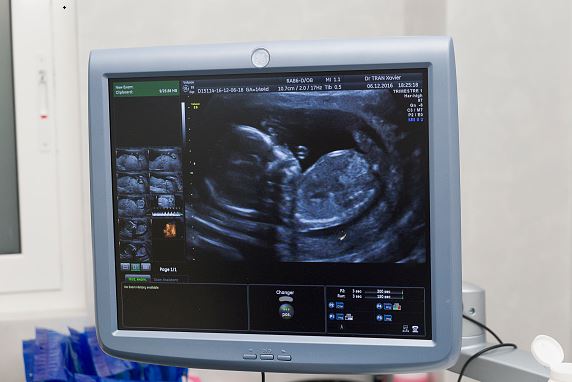 Si la placenta se encuentra en una posición inusualmente baja en la matriz, se le llama placenta previa. Generalmente esto se resuelve antes de nacer el bebé, pero en caso contrario, podría necesitar someterse a un parto por cesárea (una operación para dar a luz a su bebé a través de su barriga).
Otros ultrasonidos en el embarazo
El utrasonido puede utilizarse para otros procedimientos que se le indicarán durante el embarazo. Por ejemplo, su obstetra o ecografista pueden utilizar una ecografía para guiar una aguja fina a través del abdomen para tomar una muestra del líquido amniótico que rodea al bebé para una prueba de amniocentesis o para la obtención de muestras de tejido de la placenta para el muestreo de vellosidades coriónicas.
Es posible que le hagan otros ultrasonidos durante el embarazo si sus exámenes exploratorios de rutina o citas prenatales sugieren que pudiera haber un problema con su bebé o la placenta. Por ejemplo, le podrían hacer más ecografías si:
Ultrasonido doppler
El ultrasonido Doppler controla el flujo en los vasos sanguíneos y, en ocasiones, se utiliza para comprobar el funcionamiento de su placenta, ya que esto puede afectar el crecimiento y el desarrollo del bebé.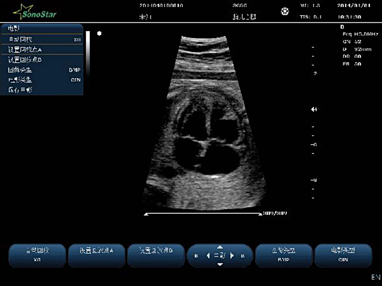 Esta no es una prueba de rutina, solo se hace si su obstetra o partera piensa que pudiera haber un problema con la placenta.
Ecocardiograma fetal
El ecocardiograma fetal es un tipo de ultrasonido Doppler realizado por un especialista para examinar el corazón de su bebé antes del nacimiento. Generalmente se hace alrededor de las semanas 18 a 23 mediante un examen exploratorio del abdomen. El ecocardiograma fetal se realiza solo si un examen exploratorio de rutina muestra anormalidades, o si su bebé tiene riesgo de tener problemas del corazón, como una cardiopatía congénita.
El médico o la partera harán los arreglos necesarios para el ultrasonido. Usted por lo regular se hará el examen exploratorio en el departamento ambulatorio de un hospital o de una clínica.
El obstetra o ecografista le explicará cómo debe prepararse para el procedimiento. Al principio del embarazo posiblemente necesite tener la vejiga llena, de modo que tendrá que beber líquidos aproximadamente una hora antes del examen. La vejiga llena ayuda a elevar el intestino grueso en la pelvis para que su matriz (el útero) se vea más fácilmente.
El obstetra o ecografista le explicará lo que sucederá antes, durante y después del tratamiento, y cualquier dolor que pueda tener. Esta es su oportunidad para entender lo que sucederá, y puede resultarle útil preparar una lista de preguntas sobre los riesgos, los beneficios y las alternativas al procedimiento. Esto le ayudará a estar informada, de modo que pueda dar su consentimiento si le piden que firme un formulario de consentimiento para llevar a cabo el procedimiento.
Un ultrasonido normalmente dura entre cinco y 15 minutos. Un ultrasonido Doppler o ecocardiograma fetal pudiera demorar más, según el tipo de investigación.
El escáner de ultrasonido se parece un poco a un sistema de computación para el hogar. Incluye un disco duro, un teclado y una pantalla. El obstetra o ecografista sostendrá un sensor que emite ondas sonoras y capta los ecos resultantes.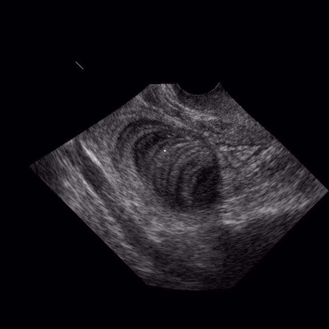 Estas imágenes se actualizan constantemente, de modo que el examen exploratorio puede mostrar los movimientos de su bebé.
Se le puede hacer el ultrasonido a través de su vagina o abdomen, dependiendo de cuántas semanas de embarazo tenga. Tanto el ultrasonido para determinar la edad gestacional como el que detecta anomalías fetales son generalmente exámenes exploratorios abdominales.
Examen exploratorio vaginal
Este método se usa si el examen exploratorio se hace en los primeros meses del embarazo (antes de aproximadamente la semana 12) cuando el embrión es muy pequeño. En esta etapa, el examen exploratorio vaginal da una vista mejor en comparación con uno abdominal.
Se le pedirá acostarse boca arriba y el médico o ecografista insertará cuidadosamente un sensor lubricado (del tamaño de un tampón) en la vagina. Generalmente el sensor se cubre con un condón. Dígale a la persona que la examine si usted padece de alergia al látex, de modo que se use un condón adecuado.
Examen exploratorio abdominal
Este método se usa normalmente en exámenes exploratorios después de la semana 10 del embarazo.
Se le pedirá que se acueste boca arriba. El ecografista u obstetra frota un gel transparente en la piel de la parte baja del abdomen. El gel permite que el sensor se deslice fácilmente sobre su piel y ayuda a producir imágenes más claras. El sensor se sostiene firmemente contra su piel y se mueve sobre la superficie. Si mira a la pantalla, verá la imagen de su bebé.
Al terminar el examen exploratorio, puede regresar a su hogar. Se conservarán copias permanentes del examen exploratorio en la computadora, en un disco o impresas. El obstetra o ecografista podría darle una copia impresa del examen exploratorio para que se la lleve después de hacerse sus exámenes de rutina. Algunos hospitales y clínicas guardan los exámenes exploratorios en un DVD para que usted se los lleve – pida más información a su obstetra o ecografista.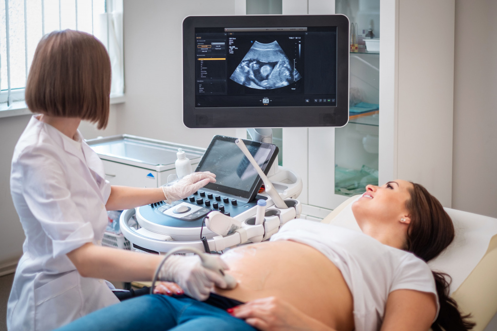
El ecografista u obstetra generalmente le explicarán los pormenores del ultrasonido durante o inmediatamente después del examen exploratorio. En ocasiones, los resultados de su sonograma se enviarán a la partera o médico que lo indicó, y usted deberá hacer una cita para conocer los resultados.
Generalmente, podrá regresar a su casa cuando sienta que está lista.
El examen de ultrasonido no causa dolor y es seguro. No emplea radiaciones ni tiene efectos dañinos sobre su bebé. Su uso se considera seguro durante el embarazo.
Un embarazo ectópico tiene que ser tratado porque pone en riesgo su vida y el embrión no sobrevivirá.
Un ultrasonido puede detectar un embarazo ectópico al menos en la semana cinco. Un embarazo ectópico es cuando el embrión se implanta fuera del útero, generalmente en una trompa de falopio, y a veces en un ovario o en el cuello del útero (cérvix).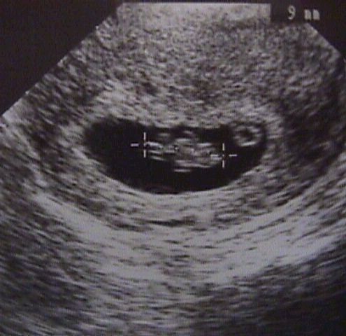
Un embrión que se implanta fuera del útero no se puede desarrollar normalmente y puede dañar el órgano al que se ha implantado, causando un sangrado abundante y poniendo en riesgo la vida de la mujer. Por ello, un embarazo ectópico confirmado necesita ser interrumpido, con el uso de medicamentos o cirugía.
La mayoría de los embarazos ectópicos se tratan usando un medicamento llamado metotrexato. Este impide que se produzcan nuevas células y de esa manera detiene el desarrollo del embarazo. El metotrexato generalmente se administra como inyección.
El embrión se puede extirpar quirúrgicamente mediante cirugía laparoscópica o abierta. En la cirugía laparoscópica, se introducen instrumentos especiales en el abdomen a través de pequeños cortes. Estos instrumentos se usan para examinar y extirpar el embarazo ectópico. En la cirugía abierta, el cirujano hace un solo corte en el abdomen y extirpa el embrión. El tratamiento del embarazo ectópico mediante cirugía es generalmente una emergencia médica.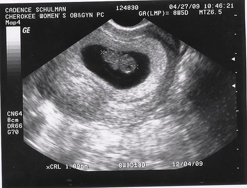
Un ultrasonido puede confirmar el sexo de su bebé en la semana 15 del embarazo aproximadamente.
Un ultrasonido puede confirmar el sexo de su bebé más o menos en el cuarto mes del embarazo. Sin embargo, no es del todo preciso porque depende de la posición de su bebé y la habilidad del auxiliar de ecografía. Usted podría averiguar en el examen exploratorio de anomalías de rutina, pero algunos hospitales no le dirán el sexo de su bebé a menos que haya un motivo médico.
Si necesita saber el sexo de su bebé por motivos médicos, se le podría ofrecer amniocéntesis o muestra de vellosidades coriónicas (MVC). Estas pruebas pueden ayudar a determinar el sexo de su bebé y detectar diversos trastornos genéticos.
La amniocéntesis implica tomar una muestra del líquido amniótico que rodea a su bebé en el útero. La amniocéntesis tiene un pequeño riesgo de causar un aborto espontáneo. Por ello solo se ofrece a mujeres cuyos exámenes de detección muestran que tienen un riesgo mayor de dar a luz un bebé con un trastorno genético, o a las mayores de 35 años de edad, ya que las posibilidaddes de tener un bebé con síndrome de Down aumentan con la edad de la madre.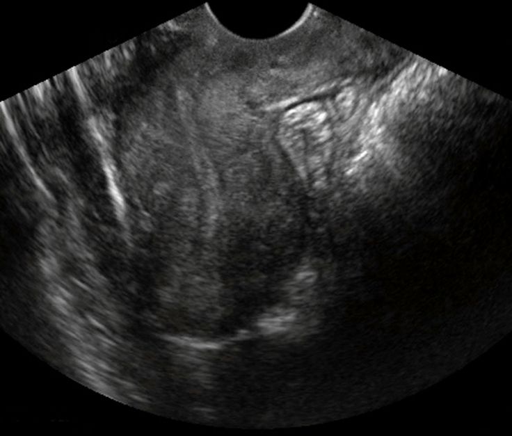
Las muestras de vellosidades coriónicas (MVC) implican la toma de pequeñas muestras de tejido de la placenta. Las muestras de vellosidades coriónicas por lo general se toman entre las semanas 10 y 13 del embarazo. El procedimiento tiene un riesgo ligeramente mayor de un aborto espontáneo en comparación con la amniocéntesis. Tampoco es tan preciso como la amniocéntesis.
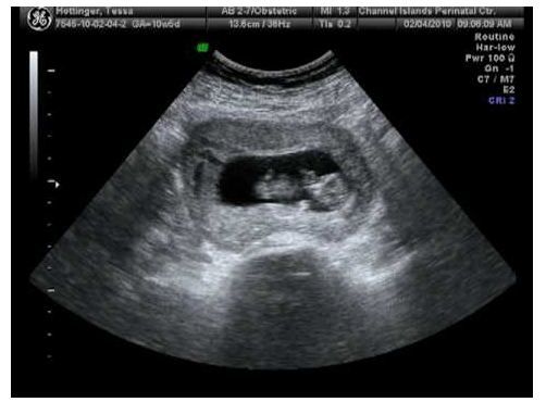
Generalmente se le hará un ultrasonido entre las semanas 10 y 14 del embarazo. Con frecuencia este se llama ultrasonido para determinar la edad gestacional porque se hace para saber cuántas semanas tiene de embarazo y estimar su posible fecha de parto. Durante este procedimiento, se podría examinar si su bebé presenta síndrome de Down.
Su partera o médico le puede decir cuán avanzado está su embarazo midiendo la longitud de su bebé de la coronilla a la rabadilla. A esto se le llama longitud céfalo-caudal (CRL). En promedio, su bebé mide de 3 a 8 cm entre las semanas 10 y 14 del embarazo.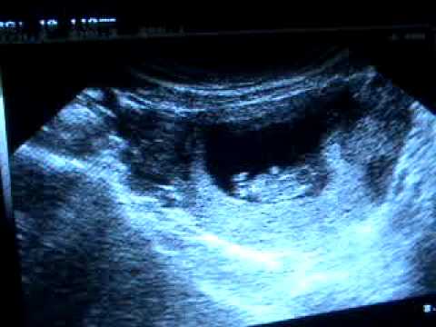 La cara de su bebé está bien formada y sus párpados están cerrados. Su bebé puede abrir y cerrar la boca y fruncir el ceño. Los brazos y las piernas son largos y delgados, y el bebé puede cerrar el puño y doblar los dedos de los pies. Los órganos sexuales de su bebé están desarrollados pero son demasiado pequeños para verse en el examen exploratorio.
Se puede medir la cantidad de líquido en un doblez detrás del cuello de su bebé para evaluar el riesgo de síndrome de Down. A esto se le llama examen de translucencia nucal. Mientras mayor sea la cantidad de líquido, mayor posibilidad de que su bebé tenga síndrome de Down. Las personas con síndrome de Down tienen un cromosoma 21 de más. Tienen características físicas y mentales típicas, tales como dificultades en el aprendizaje, rasgos faciales particulares y problemas cardíacos.
Si el examen indica un riesgo mayor, se le ofrecerá someterse a pruebas como la amniocéntesis o la muestra de vellosidades coriónicas (MVC), las cuales le pueden decir si su bebé tiene síndrome de Down.
01
Salud familiar e infantil
02
Salud familiar e infantil
TikTok
Upload
ULTRASONIDO DE LOS 8 MESES 🥰🥰🥰 #ultrasonido #ochomeses #8meses #bebe #prontonacera #embarazo #pataditas
81 Likes, 5 Comments. TikTok video from Gerardo & Aurora (@gerjuny): “ULTRASONIDO DE LOS 8 MESES 🥰🥰🥰 #ultrasonido #ochomeses #8meses #bebe #prontonacera #embarazo #pataditas”. ULTRASONIDO DE LOS 8 MESES . Original Sound.
2360 views|
#8 meses de embarazo.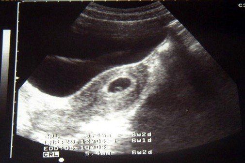 el tercer ultrasonido de mi bebé con riesgo de síndrome de down. solo Dios tiene la última palabra 🙏 🙌
TikTok video from Josefina Solano (@josefina_sinlimites): “#8 meses de embarazo. el tercer ultrasonido de mi bebé con riesgo de síndrome de down. solo Dios tiene la última palabra 🙏 🙌”. Gracias a Dios en el tercer ultrasonido mi bebé crecio normal, que no me siga preocupando del riesgo del síndrome de down.y si asi fuera yo igual lo espero con mucho amor y me siento capaz de cuidarlo y protegerlo como una leona🦁💘 . corridosybanda12.
2158 views|
We’re almost there😩🥺💕 #momanddad #babygirl #8monthspregnant #fyp #ultrasound #greenscreen
2.4K Likes, 37 Comments. TikTok video from Yazmin (@papaafritta): “We’re almost there😩🥺💕 #momanddad #babygirl #8monthspregnant #fyp #ultrasound #greenscreen”.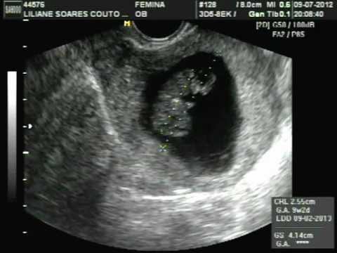 #8monthspregnant | Weighing 4lbs 8oz | Baby girl is growing big and healthy😩💕 | …. Hermoso Cariño.
32.4K views|
✨8 meses e estamos quase lá 🤰🏽✨ #tuvens #avozdoanjosusurrounomeuouvido #ultrassomnabarriga #croma #efeitoultrassombarriga #8meses #gestacao #mae
120 Likes, 11 Comments. TikTok video from Lucilene Santos (@lucilenecheck): “✨8 meses e estamos quase lá 🤰🏽✨ #tuvens #avozdoanjosusurrounomeuouvido #ultrassomnabarriga #croma #efeitoultrassombarriga #8meses #gestacao #mae”. som original.
5806 views|
Trend de embarazada💖 #8meses #embarazo #amor❤️ #pronto #viral
6. 4K Likes, 8 Comments. TikTok video from Barbara Gaytan Moren (@barbara_gaytan): “Trend de embarazada💖 #8meses #embarazo #amor❤️ #pronto #viral”. TREND DE EMBARAZADA🤰 | PRIMER PRUEBA😳😥 | PRIMER ULTRASONIDO😍💘 | …. Halo (Instrumental Version).
177.7K views|
#parati #bebestiktoks #baby
5.1K Likes, 93 Comments. TikTok video from AimeeTapiaLeon (@aimeetale): “#parati #bebestiktoks #baby”. Ahorramos para el ultrasonido 5D | Quise esperar tener los 8 meses para verlo bien | Comí un chocolate antes para que tuviera más movimiento | …. Oh No.
446.4K views|
#gestacao #menino #ultrassom
TikTok video from Fernanda Coutinho (@fercoutinho7): “#gestacao #menino #ultrassom”.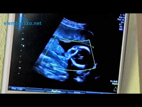 Théo 8 meses | Dia de Ultrassom
33 semanas+4
Ele já está com
2,630 kilos e com 47 cm…
Aí ai meu Deus tem mais semanas pela frente
. Photograph.
2104 views|
#parati #fyp #viral
2.1K Likes, 21 Comments. TikTok video from Raquel❤️ (@raquel2925): “#parati #fyp #viral”. tenía 16 años sali embarazada todo sentí miedo pero ala misma vez feliz | 8 meses de embarazo me hicieron un ultrasonido salió que mi hijo traía una hernia en ombligo | fui al hospital me hicieron ultrasonido y salía que el niño tenía una citometría 21 me dijeron que no iba a vivir que no me hiciera ilusiones | …. sonido original.
104.7K views|
#pov #embarazada #8mesesembarazo #fyp #ex #humor #contenido #37semanasdeembarazo #abusonarcisita #8meses
1. 7K Likes, 6 Comments. TikTok video from KARLA (@karla.vivv): “#pov #embarazada #8mesesembarazo #fyp #ex #humor #contenido #37semanasdeembarazo #abusonarcisita #8meses”. Cuando en el ultrasonido tiene la misma jeta que el papa 💩. sonido original.
36.4K views|
Uma sensacao unica ❤️ #gestante #gravidez #gravida #fy
2.6K Likes, 96 Comments. TikTok video from Julio Rocha (@juliorocha): “Uma sensacao unica ❤️ #gestante #gravidez #gravida #fy”. som original.
34K views|
During the development of the fetus, ultrasound is one of the key methods of examination. There is no exact schedule for ultrasound, so the doctor focuses primarily on the condition of the patient, the condition of the fetus, the presence of somatic pathology.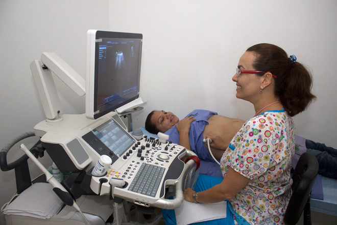 If we take a physiologically occurring pregnancy, then ultrasound is performed in each of the trimesters.
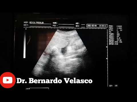
Ultrasound at 8 weeks is the first time that this study is recommended. The eighth week is the first critical period, therefore, it is most rational to carry out diagnostics during this period.
Since it is the 8th week of pregnancy, a transvaginal examination is performed more often, which is more reliable. At this time, transabdominal ultrasound is used less and less. The transvaginal method does not pose any threat to either the mother or the fetus.
If the doctor confirms a physiologically proceeding pregnancy on an ultrasound examination, does not reveal complications, the fetus develops according to age norms, then the next ultrasound is prescribed at 12 weeks.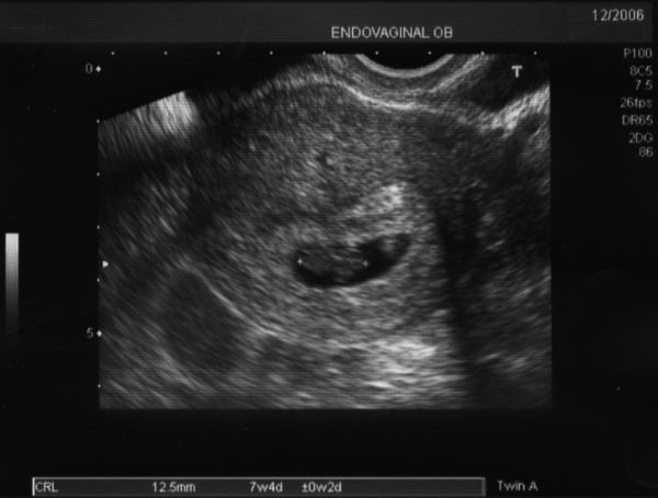 This period is another critical period when it is necessary to assess the condition of the woman and the unborn baby.
If the first two critical periods are mainly related to hormonal changes, then the next period – 22 weeks, is most often associated with sexual infections. Therefore, we also perform an ultrasound at 22 weeks. Pregnancy can provoke the development of latent infections, which, in turn, can lead to certain complications.
There are no exact recommendations for registering pregnant women, but doctors recommend starting monitoring at the antenatal clinic before 12 weeks. There is a mandatory list of tests in accordance with the order of the Ministry of Health of the Russian Federation, and an additional one, which is appointed by the gynecologist individually.
Tests at 8 weeks of gestation:
In addition, the 8th week of pregnancy is the period of visiting the following doctors:
Accordingly, if any pathology is detected, any of these specialists has the right to prescribe an additional analysis in their direction. If this pregnancy proceeds with complications, or if there are previous complications in the anamnesis, then the laboratory and instrumental complex may expand.
You will have to take tests regularly, so you should prepare in advance for the next tests at week 12.
As we mentioned earlier, the next critical period is the 12th week of pregnancy. Therefore, the next ultrasound is performed at this time. Here we are not talking about a simple study, but about screening, which allows you to identify intrauterine malformations of the fetus.
In contrast to the ultrasound at 8 weeks, this is predominantly a transabdominal examination.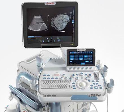 Transvaginal ultrasound is performed only for certain indications:
So, what information does an ultrasound give a gynecologist?
It must be clearly understood that ultrasound during pregnancy is only one of the methods of examination, therefore, if there are any deviations in the indicators, you should not panic, you should consult a specialist.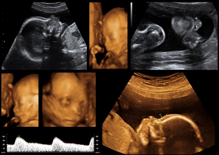 The doctor, interpreting the results, always relies on many factors, which allows him to adequately assess the true state of the future mother and fetus.
Even if deviations in some indicators are confirmed, doctors still only talk about a certain probability of developing a particular pathology. In this case, a second screening may be prescribed, or a similar study is carried out at a later date. In any case, the final decision regarding the continuation of the pregnancy is made exclusively by a specialist.
As we have already said, 12 weeks is the time for screening. For this, an ultrasound examination is prescribed, the external indicators of the woman are evaluated, and a blood test is performed.
Particular attention is paid to two coefficients: β-hCG and PAPP-A. It is believed that in the presence of deviations according to these parameters, one can judge the presence of fetal pathologies. According to the level of hCG, doctors predict the further management of pregnancy and the possibility of miscarriage or premature birth.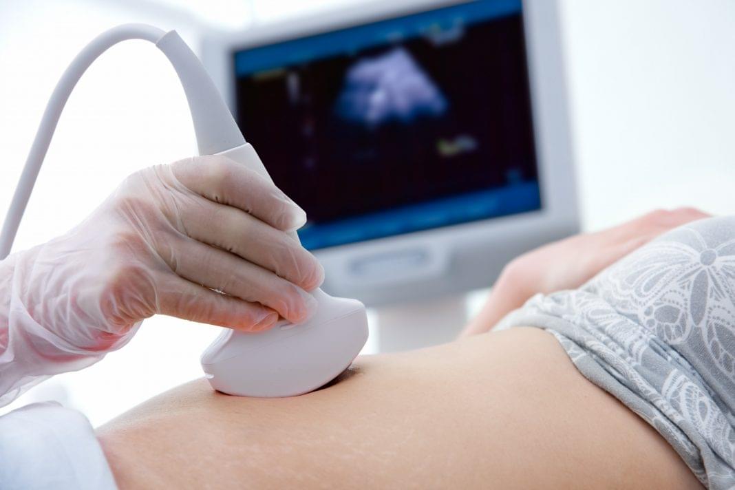
Accordingly, according to the screening results, it is possible to identify:
Thus, tests at the 12th week of pregnancy are decisive for solving a number of issues related to the further development of the fetus. It should be clearly understood that screening is not 100% reliable, therefore, if you receive any deviations from normal results, you should immediately contact a specialist who can correctly assess such changes in indicators.
The next scheduled ultrasound is performed at 22 weeks of gestation. Here, for the doctor, it is not the condition of the mother or the size of the fetus that is important, but the assessment of the formation of various organs and systems, as well as bones.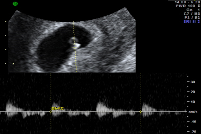
So, the 22nd week – the fetus has already formed all the main organs and systems, and on the monitor screen the child acquires the outlines familiar to future parents. Accordingly, the following indicators come to the fore:
Unlike ultrasound at 8 weeks, ultrasound at this time is performed transabdominally. 22 weeks is already the second trimester, and right now the question is being decided whether there are serious deviations in the development of the fetus, and further actions of obstetricians. The question of whether to continue a further pregnancy is decided not by one doctor, but by a whole council, based on numerous research results.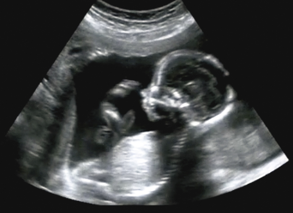
Week 22 is when the second screening is more common. This analysis is prescribed in the period from 18 to 24 weeks, but this time is considered optimal. Accordingly, during repeated screening, the doctor already makes final conclusions about the condition and development of the fetus, the presence of deviations.
In addition to ultrasound, the doctor prescribes mandatory tests to monitor the woman’s health. The expectant mother again takes a general blood and urine test, where the most important are a number of indicators: hemoglobin, erythrocytes, leukocytes, ESR, protein in the urine.
During the examination, the gynecologist necessarily measures the woman’s height, weight, blood pressure level, and measures the abdomen. Tests at 22 weeks of gestation are designed to assess the likelihood of developing preeclampsia. Here, the role is played not by hormonal changes, but by the exacerbation of chronic pathology. Therefore, timely screening and assessment of the woman’s condition allows you to plan further tactics for managing pregnancy.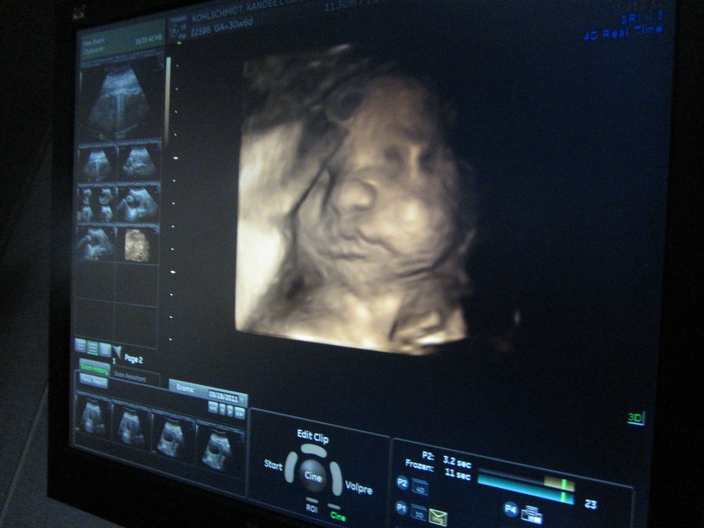
You can find out the cost of an appointment with a gynecologist by phone number +7 495 478 10 03. Also during the planning of pregnancy in our center there is an opportunity to undergo a comprehensive examination of the body for women, and for future fathers – a comprehensive examination of men.
The author of the text: Sokolov Alexander Aleksandrovich
Position: Ultrasound diagnostic doctor
Total experience: 6 years
90,000 conducting an ultrasound at 8 weeks0186
For ultrasound in the first trimester of , certain terms are set – 10-13 weeks. During the gestation period of this particular duration, the fetus will already begin laying the main internal organs, the skeleton, and the brain. With the help of ultrasound, all these parameters can be seen and evaluated.
However, it happens that the doctor prescribes an ultrasound at the 8th week of pregnancy, when it is too early to judge the presence or absence of pathologies in the fetus.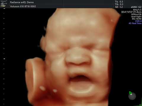 Such early research has goals.
Ultrasound at the eighth week is done for:
By the eighth week, the fetus reaches a length of two centimeters. He has already formed the circulatory system, the first veins and the main internal organs, which will continue to develop throughout the entire period of pregnancy.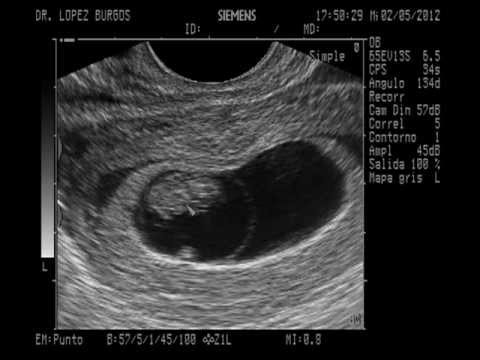 The fetal heart is already beating, and this can be seen if an ultrasound is performed.
Fingers are already appearing on the hands and feet of the baby, and the upper limbs are increasing in length. Shoulders and knees appeared. The inner ear, that is, the center of balance, begins to form.
The eyes of the fetus are forming, they are already closed by thin eyelids.
Ultrasound at the 8th obstetric week of pregnancy does not require special preparation. Women are simply advised 2-3 days before the study to refrain from food that provokes excessive gas formation in the intestines, as well as from carbonated drinks. You need to come to the doctor’s appointment calm and in a good mood.
At the eighth week, ultrasound can be performed in two ways:
The first method will give more accurate results, but may cause some discomfort to the woman. In addition, transvaginal ultrasound is not recommended if a woman complains of abdominal pain or bloody discharge.
After the ultrasound, the doctor may take a photo showing the tiny embryo.
Ultrasound at the eighth week of pregnancy, with its correct course, visualizes a fetal egg with a live embryo inside. Also, according to the results of the study, it will be possible to judge the place of attachment of the chorion, the position of the fetal egg in the uterine cavity.
Ultrasound at 8 weeks should show the fetal heartbeat and the frequency can be assessed.
At a period of 8 weeks, it is already possible to identify some pathologies in the development of the fetus, in particular in the work of his heart. At the same time, specialists have not yet made unambiguous conclusions about the presence of heart disease.
Ultrasound will also show how the neural tube of the fetus develops, how limbs are formed.
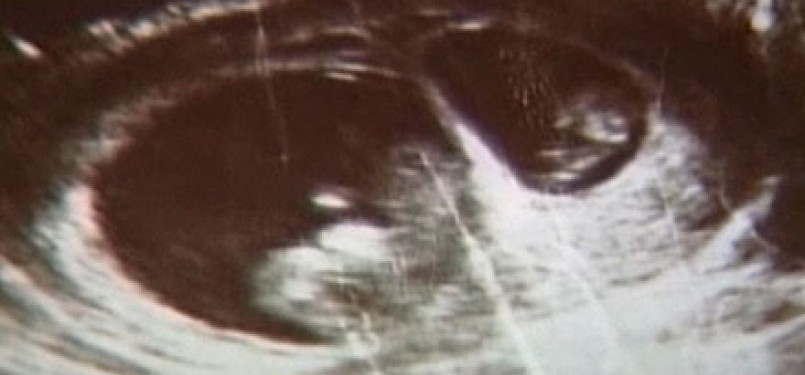
A large proportion of the total ultrasounds at the eighth week of is the study of the embryo. Its size, location, structure is important. It is also necessary to evaluate the motor activity of the embryo, which should be present even at such a short time. At the eighth week, the embryo should be clearly visible. If it is not there, most likely its development has froze or there is an anembryonic fact, that is, the initial absence of an embryo in a fetal egg (or its death immediately after conception).
When conducting an ultrasound scan at 8-9 obstetric weeks of pregnancy, it is required to assess not only the state of the embryo itself, but also the organs outside its structure. These organs include the yolk sac.
Its presence in the ovum is temporary. The pouch appears approximately 15-16 days after fertilization and disappears by the end of the first trimester.
The yolk sac plays an important role in the development of the embryo – it produces germ cells, forms embryonic red blood cells for the first time, produces proteins necessary for development, helps to form immunity, and so on.
At the eighth week, the ultrasound should show the yolk sac. Its size should be approximately 4-5 mm. An increase in the size of the yolk sac can be the first sign of pathological development of the fetus.
At the eighth week of gestation, an ultrasound is done and they look at how the parameters of the developing fetus correlate with the norm for a particular gestational age. The main indicators of fetal development include:
A deviation from the norm of one or several parameters at once can be a signal that the fetus is developing with pathology. Although doctors do not make unambiguous conclusions on such a short period of time and, as a rule, prescribe a second ultrasound in 1-2 weeks.
Book a diagnostic or consultation today!
You can make an appointment by phone:
+7 (812) 901-03-03
Or leave a request
full name
Phone number
By clicking the “Make an appointment” button, I accept the terms of the Personal Data Processing and Security Policy and consent to the processing of my personal data.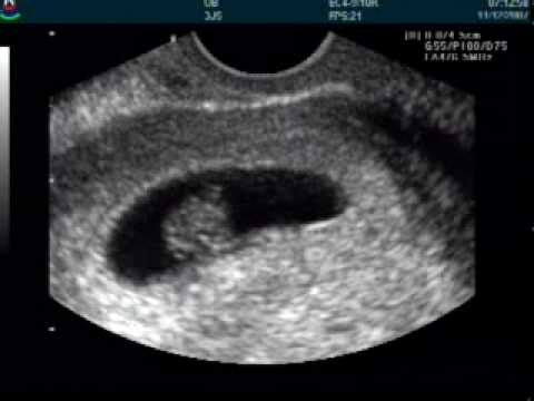
Making an appointment
Patient’s last name *
Incorrect first name
Name *
Middle name
Contact phone *
E-mail *
By clicking the “Make an appointment” button, I accept the terms of the Personal Data Processing and Security Policy and consent to the processing of my personal data.
Registration and payment for repeated online appointment
Patient’s last name *
Incorrect first name
Name *
Middle name *
Contact phone *
E-mail *
By clicking the “Submit request” button, I accept the terms of the Personal Data Processing and Security Policy and consent to the processing of my personal data.